
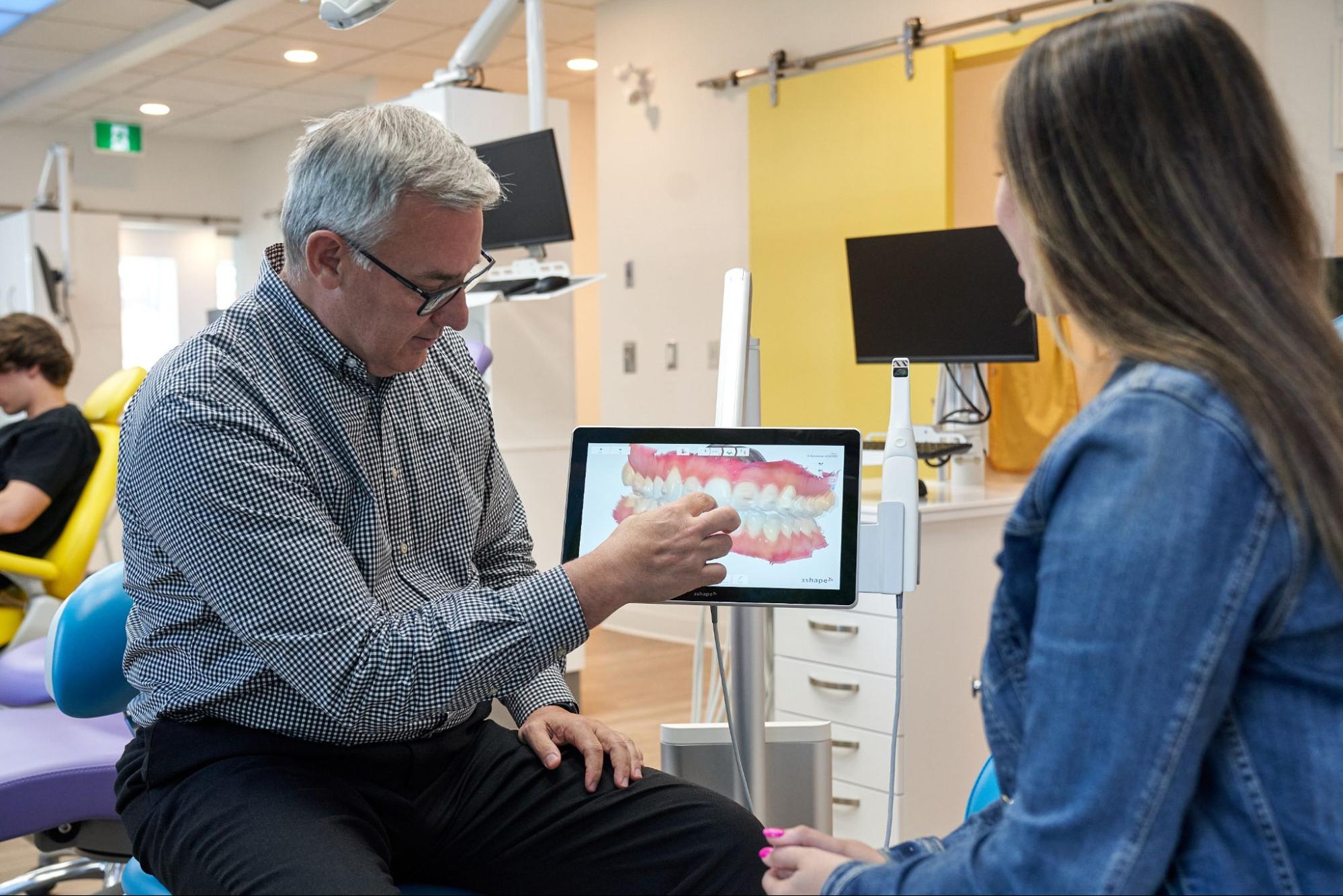
Short answer up front: what your digital scan actually shows you is a detailed 3D model of your teeth, gums, and bite that you and your doctor can rotate, zoom, and measure together on a screen. No trays. No putty. Just a few minutes with a small wand.
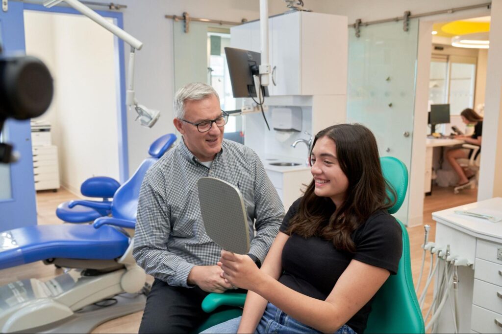
What Is a Digital Dental Scan?
A digital dental scan uses a small handheld wand to capture thousands of images of your teeth and gums, which software stitches into an accurate 3D model on screen. There’s no putty and no tray. The scan takes a few minutes per arch, uses light instead of radiation, and becomes the digital file your doctor plans treatment from.
The wand reads surface anatomy: the shape of every tooth, the height of your gum line, the way your upper and lower arches fit together. That’s a different job than an X-ray or a CBCT scan, which look through tissue at roots, bone, unerupted teeth, sinuses, and airway space.
Both matter. Neither replaces the other.
What happens to the file afterward?
Once the model is finished, it saves as a standard 3D file (usually an STL or PLY). That single file can be sent to an aligner lab, used to print a retainer, shared with a specialist, or stored as a baseline for comparison years down the road.
Because it’s digital, nothing warps in a shipping box and nothing sits in a drawer collecting dust. If a lab needs another copy, we resend it in seconds.
As for the hardware itself, families sometimes recognize brand names from other offices or from ads. The common intraoral systems include:
- iTero (often paired with clear aligner workflows)
- 3Shape TRIOS (full-color scanning)
- Primescan (high-resolution capture)
They differ in software and speed, but the principle is the same across all of them: optical sensors, structured light, and real-time model building.
How long does a scan take?
Most arches scan in about three to five minutes. Total chair time is usually under ten minutes, including the bite registration.
Kids often find it easier than adults expect, since there’s nothing filling their mouth and nothing to hold still for. Many watch their own teeth build on the monitor while it happens.
And to answer the question parents ask most: scanning is radiation-free. The wand emits light, not radiation, so there’s no dose to count and no lead apron required. That’s one reason digital scans work well for progress checks, where we may want new records more than once during a smile transformation.
How a Digital Scan Works, Step by Step
A digital scan works in six quick steps: your team dries the teeth and places a cheek retractor, the wand sweeps the upper arch, then the lower arch, a bite scan records how your teeth meet, software builds the 3D model in real time, and your doctor reviews the finished model with you chairside. Most visits wrap up in under ten minutes.
Here’s what each step looks like from the chair.
- Quick prep. We brush away loose debris and dry the teeth with a puff of air. A soft cheek retractor holds the lips out of the way so the camera has a clear view of every surface. Nothing sharp, nothing tight.
- Scanning the upper arch. The wand glides along the biting surfaces first, then the outside (buccal) surfaces, then the inside (lingual) surfaces. It never has to press hard on the gums, and it pauses whenever you need a break.
- Scanning the lower arch. Same sweep, same order. Kids who tire out can stop between arches and pick right back up, since the software holds everything already captured.
- Bite registration. You close naturally while the wand records both arches together from the side. This is the piece that tells us how the upper and lower teeth actually meet, which drives almost every decision in orthodontics.
- Real-time model building. Frames stitch together on the monitor as we go, in full color. By the time we set the wand down, the finished 3D model is already spinning on the screen next to you.
- Chairside review. Your doctor rotates the model, zooms in on specific teeth, and points out what stood out. Crowding, wear, gum levels, the midline. You see it at the same moment we do.
What if something needs a second pass?
Missed a spot? A little saliva blurred one molar? We rescan that small area in a few seconds and the software drops the clean data into place.
That’s a real advantage over older methods. With putty, a flawed record meant starting the whole tray over. With digital scans, we fix small errors on the spot and move on, which keeps the visit short and keeps kids comfortable.
What the Scan Actually Reveals: 8 Things You Can See on Screen
The model isn’t just a pretty picture. It’s a measurable record, and most of what we look at is visible to you too once someone points it out. Here are eight things worth asking about while the model is still on the screen.
- Crowding and spacing, in millimeters. Software measures the difference between the space your teeth need and the space the arch actually has. Instead of “a little crowded,” you get an actual number you can compare against later scans.
- How your bite fits together. The bite registration shows overbite, overjet, crossbite, open bite, and midline shift from angles you can’t see in a mirror. Parents are often surprised at how much a midline shift stands out on a 3D model.
- Wear facets and chipped edges. Grinding and clenching leave classic flat spots on canines and incisors. On screen, worn areas show up as shiny, flat planes where a natural curve should be.
- Gum recession and gingival margin height. Ever wonder why one tooth looks longer than its neighbour? The model maps where the gum line sits on every tooth, and uneven margins often explain it while giving us a baseline to watch over time.
- Enamel cracks and worn contact points. Craze lines, thinning edges, and spots where two teeth rub against each other become obvious under magnification. These often point to a bite issue rather than a brushing issue.
- Simulated outcome previews. Aligner software can project where teeth would sit after straightening smiles, so you see a proposed before-and-after side by side. Treat it as a planning tool rather than a promise, since biology and wear time both influence the real result.
- Erupting permanent teeth and available space (for kids). We can see arch width, where permanent teeth are pushing through, and whether there’s room for the ones still coming. That’s a big part of how we know your child’s smile is developing on schedule.
- Change over time. Compare a new scan to an older one and shifting, wear, and recession show up as measurable differences instead of impressions from memory. This matters for retainer checks, growth tracking, and anyone who grinds at night.
One honest limit: a surface scan can flag signs of trouble, but it can’t see between teeth or under the gums. Decay hiding between molars still needs radiographs to confirm.
Digital Scan vs. Putty Impression vs. X-ray: What Each One Sees
Different records answer different questions. A scan captures shape. An X-ray captures structure. Putty captures shape too, just less comfortably and with more ways to go wrong on the way to the lab.
| Digital scan | Putty impression | X-ray / CBCT | |
|---|---|---|---|
| What it captures | Tooth and gum surfaces, bite relationship | Tooth and gum surfaces only | Roots, bone, unerupted teeth, sinuses, airway |
| Comfort | No trays, no material in the mouth, low gag reflex risk | Full tray plus setting material; harder for sensitive kids | Quick and non-contact |
| Chair time | About 3-5 minutes per arch | Several minutes per arch, plus setting time | Seconds to a few minutes |
| Radiation | None (optical light only) | None | Low-dose radiation |
| Storage and reuse | Digital file, resent instantly, stored indefinitely | Single-use physical model, can distort in transit | Digital image stored in your chart |
| Best for | Aligners, retainers, nightguards, progress tracking | Backup when scanning isn’t possible | Diagnosing what sits below the surface |
Do these records work together?
They do. A scan tells us where the crowns are today. A panoramic film or CBCT tells us what the roots are doing, whether a permanent tooth is stuck, and how the jaws are built. Plan a smile transformation from only one of those and you’re working half-blind.
For most families, the records visit includes both: a digital scan for the model, plus the imaging your doctor decides is clinically necessary.
Why does the digital file keep earning its keep?
Lost a retainer six months after treatment? If we still have your scan on file, we can often print a replacement from the stored model instead of starting from scratch. Older putty models simply couldn’t do that.
Records also travel well. If you move, need a second opinion, or see a specialist, the file transfers cleanly.
Who Should Get a Digital Scan (and When)
Scanning fits almost everyone, but the timing tends to cluster around a few specific moments.
- Kids around age 7. The American Association of Orthodontists recommends a first orthodontic evaluation by age 7, when enough permanent teeth have arrived to show how the bite is developing. That window is early enough to catch a developing bite issue and late enough that the permanent teeth have started to show their pattern. A scan at that visit creates a baseline we can measure against for years.
- Teens and adults considering treatment. If you’re weighing clear aligners, metal braces, or clear braces, the scan is the starting point for planning and for seeing a projected outcome.
- Anyone with a strong gag reflex. Old-style trays were rough on sensitive kids and adults alike. A wand solves that, because nothing sits in the mouth and you can pause any time.
- Families needing a replacement retainer or a nightguard. A fresh scan produces a precise appliance without a lab tray, and the stored file makes future replacements easier.
- Anyone tracking change. Grinding, gum recession, and slow shifting are best judged by comparison, not memory. Repeat scans give us that comparison.
When is scanning not the right tool?
Very limited mouth opening, certain oral appliances, or difficulty sitting still for a few minutes can make scanning tough. Some kids with sensory sensitivities do better with a shorter, staged approach across two visits.
We work around those situations rather than force them. Dr. Adriana Salles, our pediatric dentist, often breaks the scan into small sections with breaks between so younger kids stay comfortable from start to finish.
Frequently Asked Questions About Digital Scans
Does a digital scan hurt?
No. There are no trays, no putty, and nothing pressing against the gums or palate. The wand is about the size of an electric toothbrush head and only touches teeth lightly as it moves. Most people describe it as mildly ticklish at worst.
How long does a digital scan take?
Usually three to five minutes per arch, with total chair time under ten minutes including the bite scan. Kids who need breaks can take them without losing any of the data already captured. If a small area needs a second pass, the rescan takes seconds.
Is there radiation from a digital scan?
No. Intraoral scanners use light and optical sensors to map surfaces, so there’s no radiation dose involved. That’s different from X-rays and CBCT, which use low-dose radiation to see roots, bone, and unerupted teeth. Because scanning is radiation-free, repeat scans for progress tracking are simple to take.
Can a digital scan detect cavities?
It can show surface signs like discoloration, rough spots, and breakdown on visible tooth surfaces. What it can’t do is see between teeth or beneath existing fillings, which is where many cavities start. X-rays remain necessary for a full diagnosis, and your doctor will combine both when deciding on treatment.
Can I see my scan results the same day?
Yes. The 3D model builds on the monitor in real time, so it’s finished before you leave the chair. Your doctor can rotate it, zoom in, and walk you through specific findings during the same visit. Many families take a photo of the screen to show the rest of the household.
Are digital scans accurate enough for aligners?
Yes. Digital scans are the standard input for clear aligner fabrication and for printed retainers, and labs build appliances directly from the file. Accuracy depends on good technique, which is why we dry the teeth, use retraction, and rescan any area that looks unclear before we finish.
Seeing Your Own Smile Makes the Plan Easier to Trust
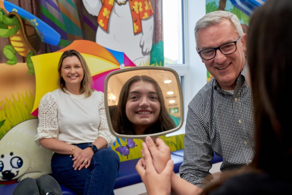
There’s a real difference between being told what’s happening in your mouth and watching it spin on a screen in front of you. That’s the part families remember most about digital scans.
Topsmiles Pediatric Dentistry & Orthodontics is a dual-specialty office, which means kids dentistry and orthodontics happen under one roof, from birth to braces. Dr. Alvaro Salles handles orthodontic treatment and Dr. Adriana Salles leads pediatric dental care, and they review scan records together when a case calls for it.
Our two offices welcome new families, and you’re welcome to ask questions about your model for as long as you’d like while it’s on the screen. If a free ortho consult is where you’re starting, bring those questions with you.
You can see your own model at your first visit. Ask the team to keep it on the monitor and walk you through it tooth by tooth, arch by arch, so you leave knowing what the plan is built on. That’s where the plan starts to make sense, and where you begin to discover your top smile with a team building toward a lifetime of healthy smiles.